Mouse over the image
Mouse over the left image
Mouse over the left image
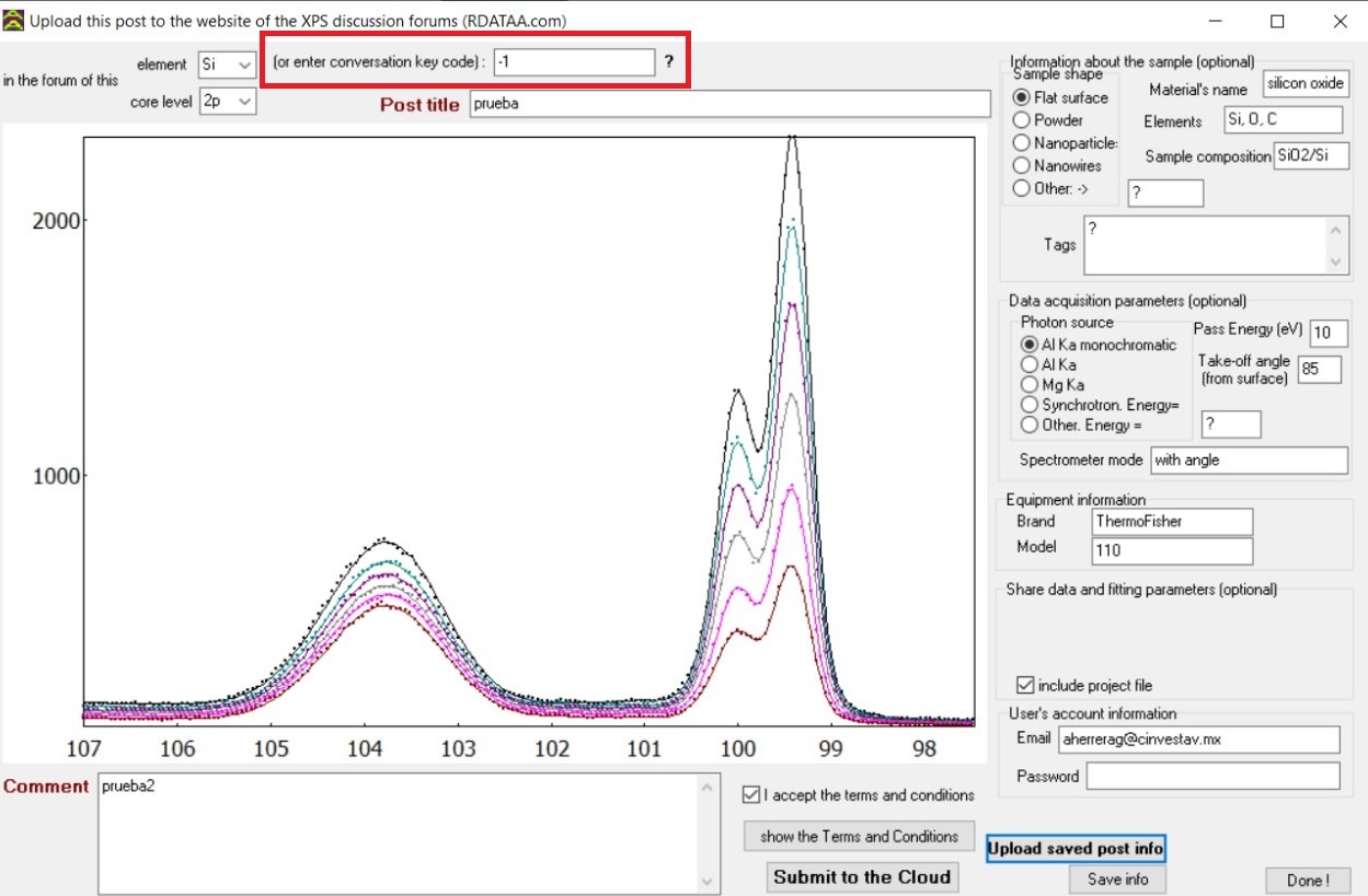
You can use it to post a comment from AAnalyzer by pasting it in the region shown below.
Number of posts created
Most voted posts
More related groups
Users with more verified posts
Do you have a question about your fit? Get help by posting it and waiting for suggestions from other users!
Do you want to inspect fits made by other users and see how they are struggling? Pick a core level and join the discussion!
Do you have suggestions? Go ahead and help other users!
Do you believe that a set of posted peak-fitting parameters might be useful to you? Download it and apply them to your data!
Is the forum of your interest still empty? Be the first one to post!


