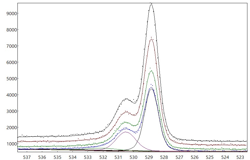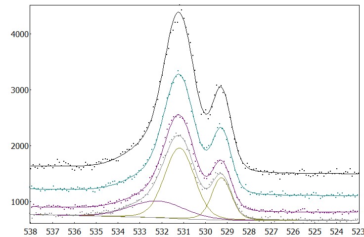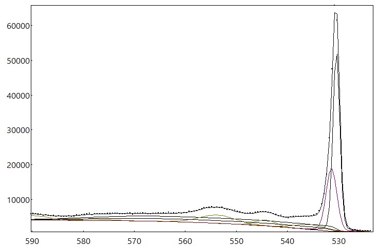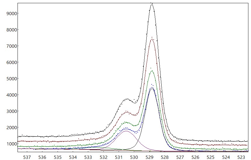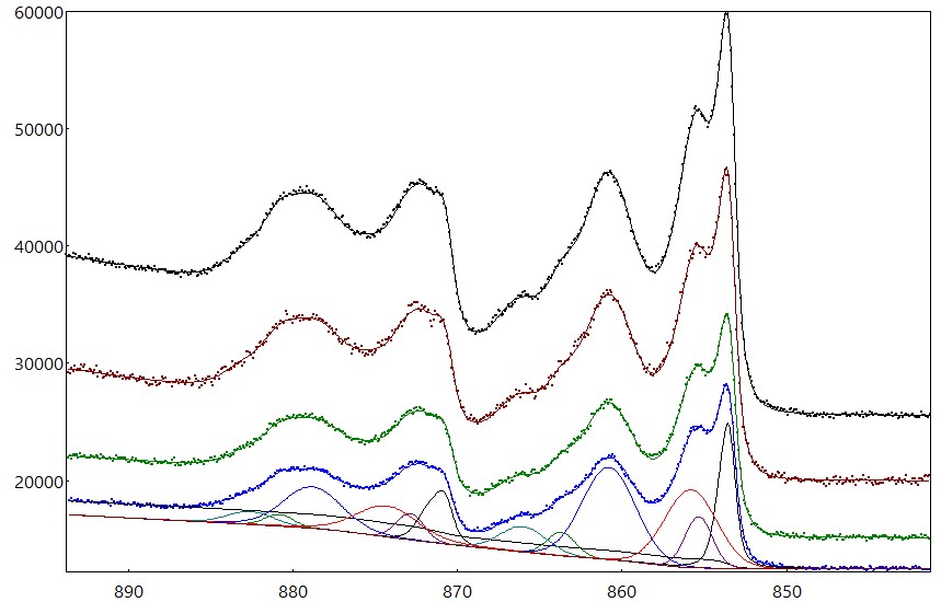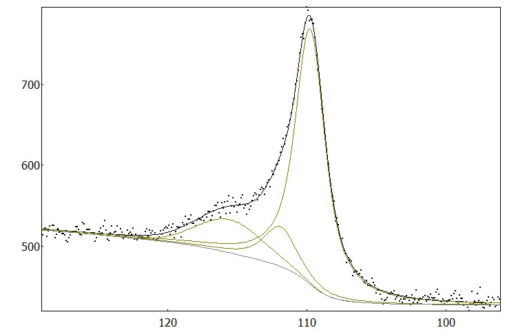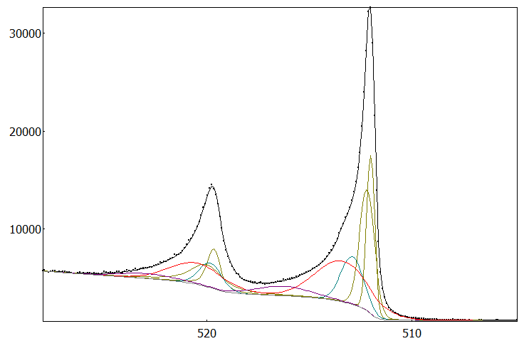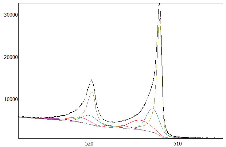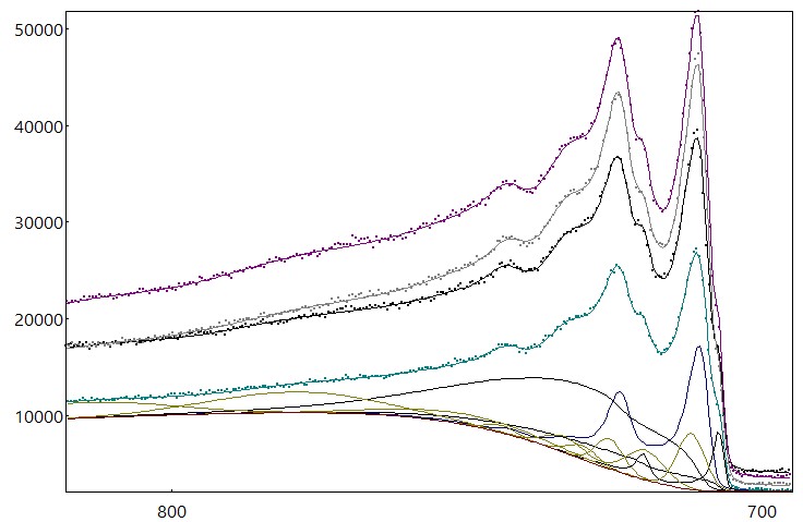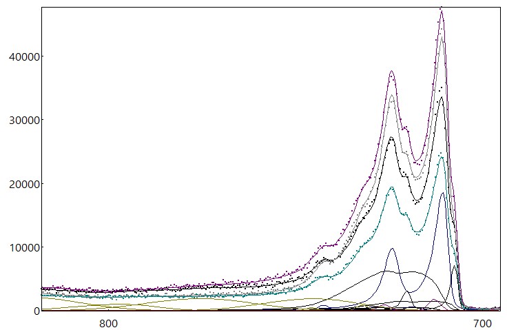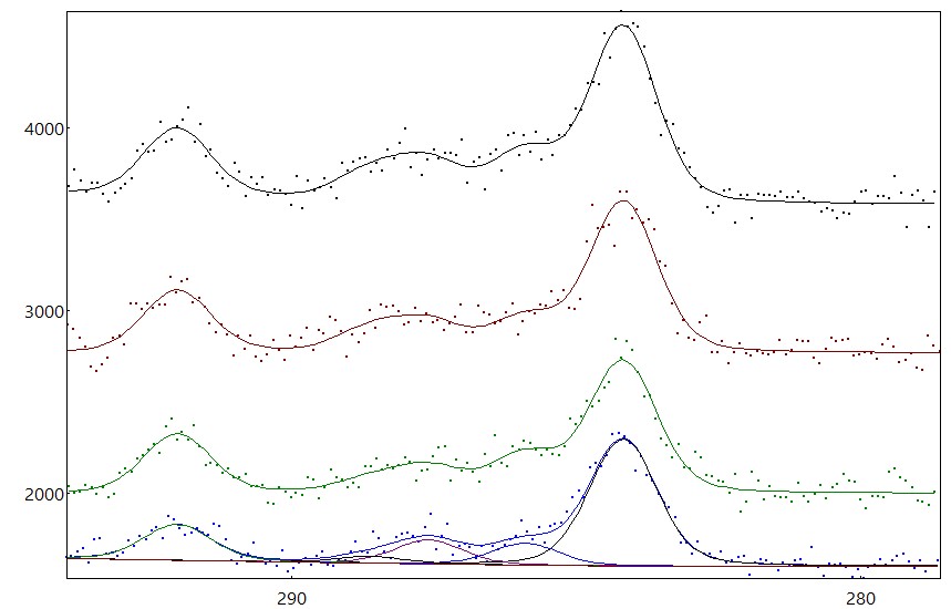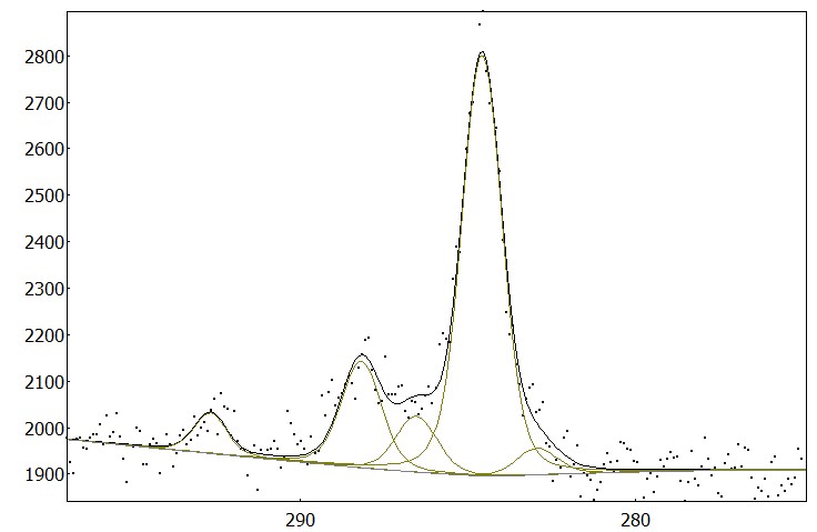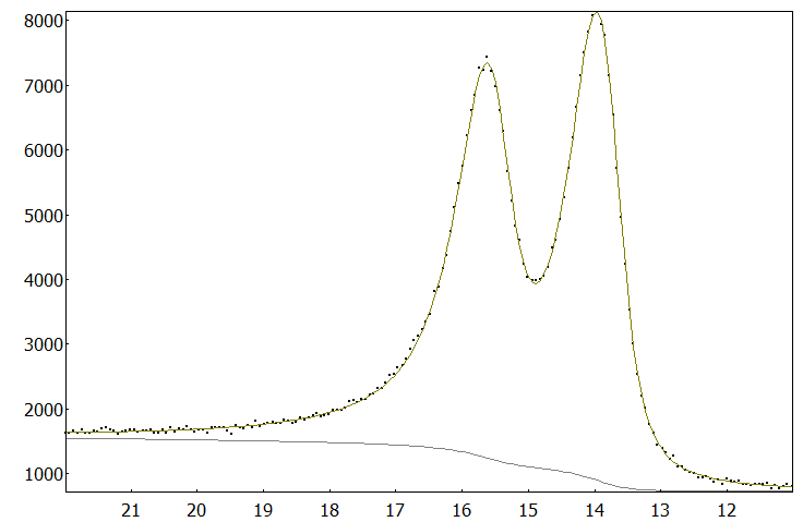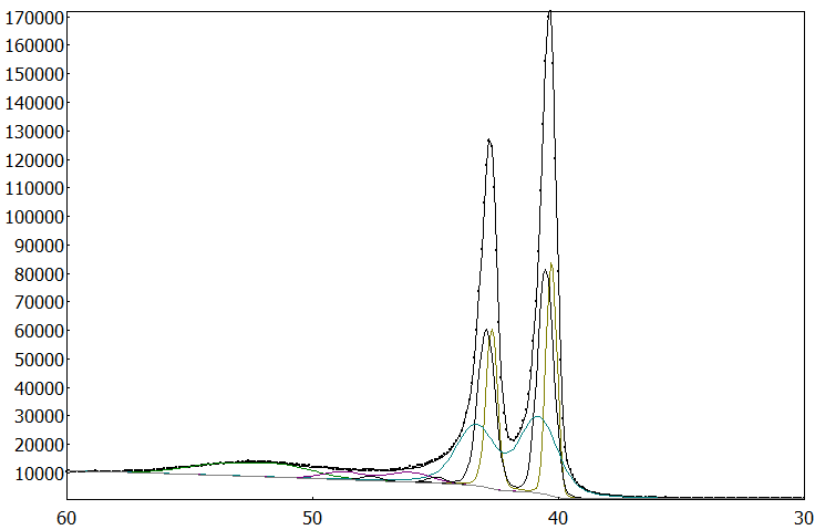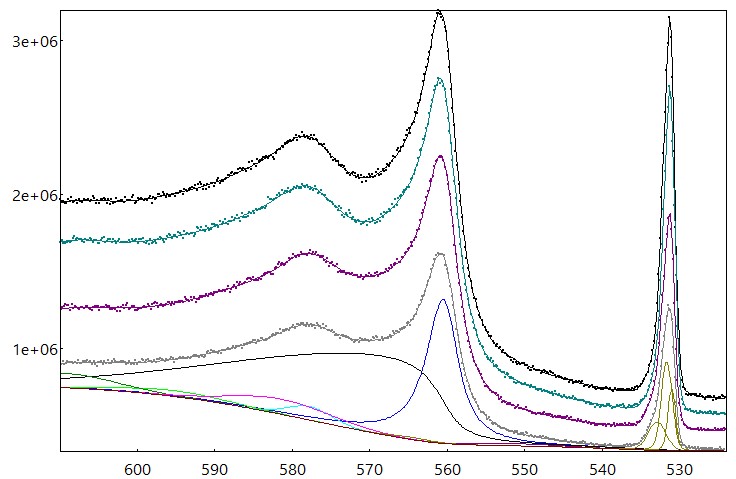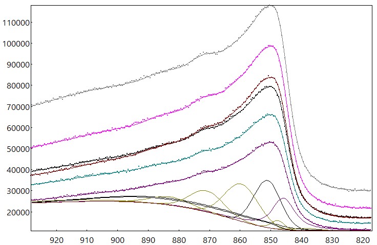
O 1s spectra was obtained from the surface of a hafnium oxide sample that was deposited by atomic layer deposition over Si(1 0 0) wafers. The component at ~530 eV is associated to Hf-O bonding in hafnium oxide. The component at 532 eV has contributions from the oxygen of the superficial hydroxil groups and from the oxygen in hafnium silicate at the interface HfO2/Si.
Read moreShape: Flat surface
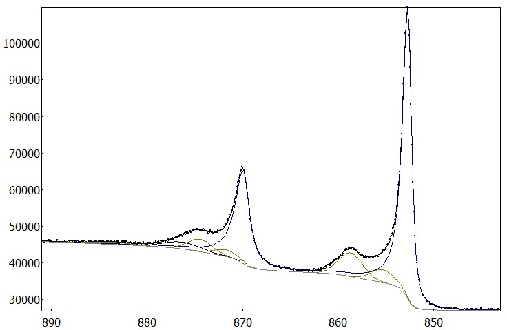
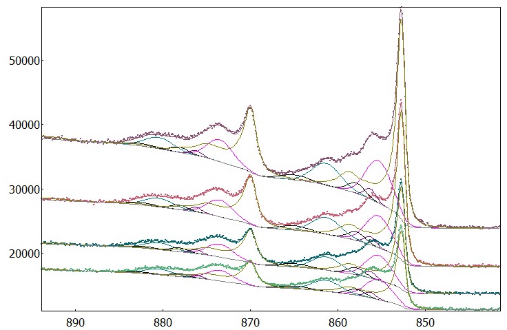
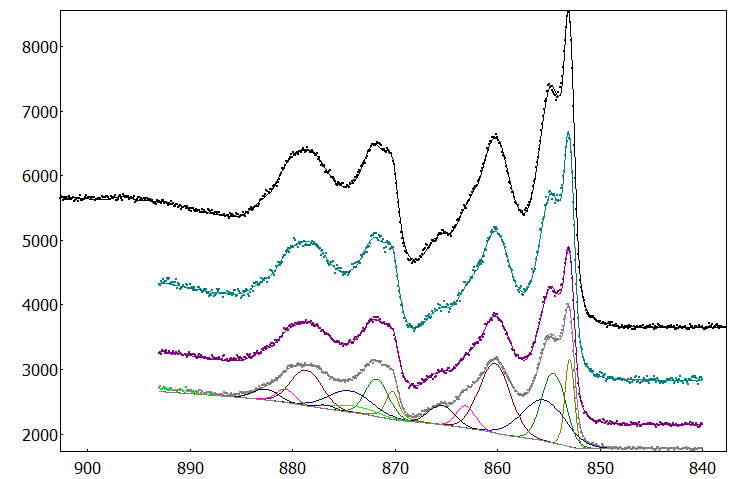
Sample material: Hafnium oxide
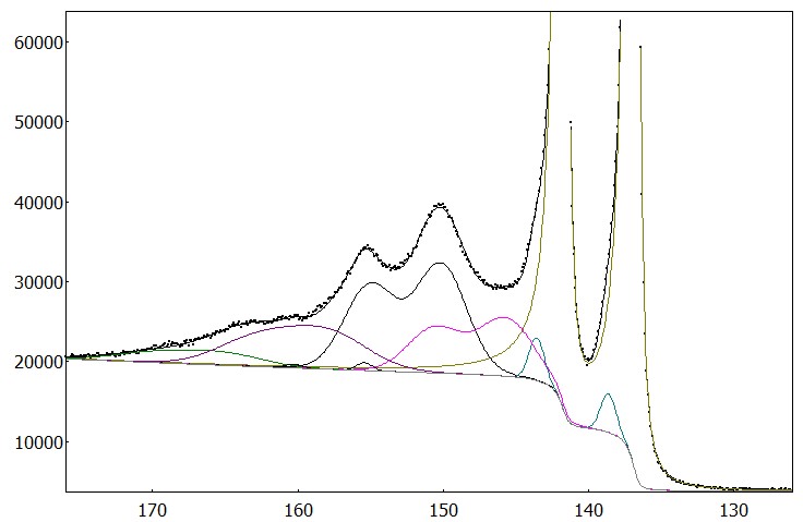
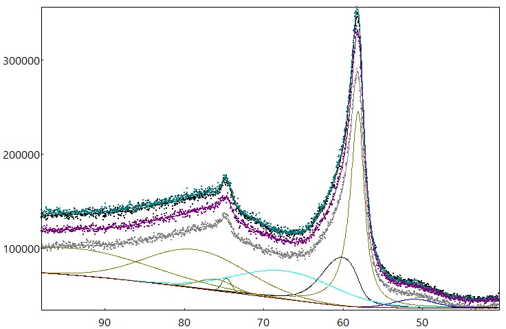
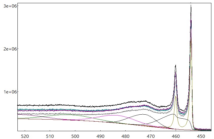
Elements: Hf, O, Si
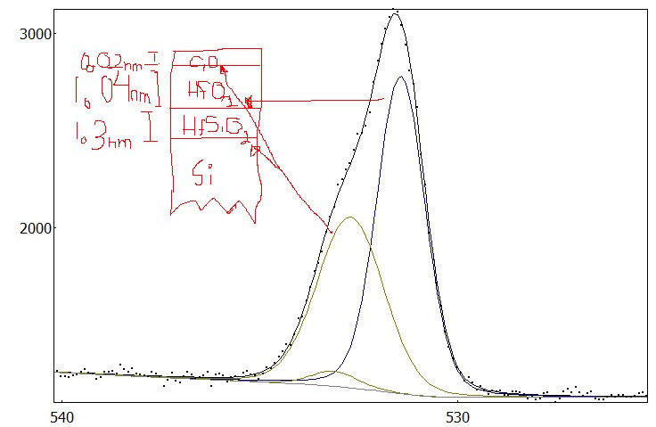
Sample composition: HfO2/HfSiO2/Si
Photon source: Al Ka monochromatic
Take-off angle: 85°
Brand: ThermoFisher
Model: Alpha 110
Spectrometer mode: large angle, small area